Pacchetto Confocale
Interfaccia dedicata per i sistemi confocali e multifotone di Nikon, che offre una facile configurazione dello strumento e operazioni semplificate. Incorpora molte delle caratteristiche di NIS-Elements AR per acquisizione avanzata, elaborazione delle immagini, analisi, visualizzazione e capacità di condivisione dei dati.
Caratteristiche principali
Enhance your Confocal Resolution with ER
Higher resolution confocal images can be easily generated with a single click. The software assesses the captured image and automatically determines processing parameters to achieve enhanced resolution.
ER can be applied to previously captured confocal images as well.
Resolution
Image resolution is defined as the smallest distance between 2 points that can be resolved. The theoretical limit of resolution for a conventional optical microscope is approx. 200nm. Higher resolution images can be theoretically achieved with confocal microscopes, but this has not been effectively achieved. Using unique image processing technology, image resolution can be increased beyond that of a conventional confocal image (~ 1.5 times improvement in XY; ~1.7 times improvement in Z).

Standard confocal image

Enhanced Resolution image
Zebrafish lens at 5 dpf. Nuclei (green) and actin filaments (red) are visualized with Cytox-green and Rhadamine-conjugated phalloidin, respectively. High magnification images indicate lens fiber cells, which become flat and stack against each other.
Image courtesy of: Drs. Toshiaki Mochizuki and Ichiro Masai, Developmental Neurobiology Unit, Okinawa Institute of Science and Technology Graduate University

Standard confocal image
Enhanced Resolution image

Standard confocal image

Enhanced Resolution image
Stress Fibers (LLC-PK1, Pig Kidney Cells), Green: F-actin, Red: Myosin Heavy Chain
Image courtesy of: Keiju Kamijo Ph.D., Division of Anatomy and Cell Biology, Faculty of Medicine, Tohoku Medical and Pharmaceutical University
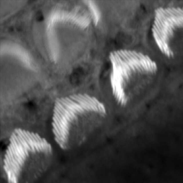
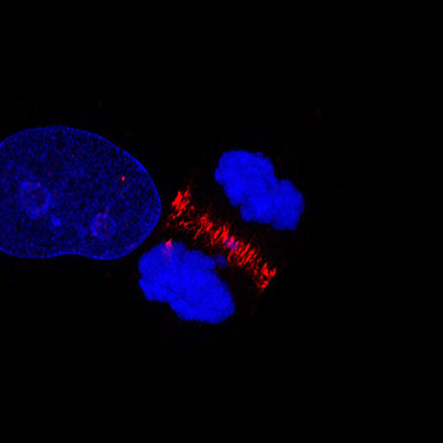
Standard confocal image

Enhanced Resolution image
Apical surfaces of auditory epithelia of mouse cochleae were stained by Atto-565-phalloidin at postnatal day 2.
Image courtesy of: Dr. Hideru Togashi, Division of Molecular and Cellular Biology, Department of Biochemistry and Molecular Biology, Kobe University Graduate School of Medicine
Upgradability
Enhanced Resolution mode is compatible with Nikon confocal microscopes. An upgrade plan is available for current NIS-Elements C users.
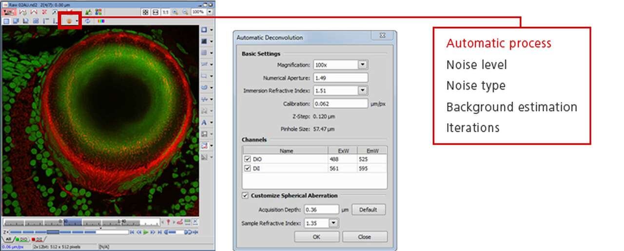
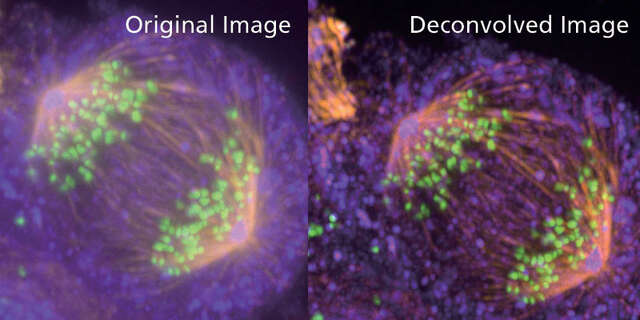
See for yourself…test our deconvolution for free!
NIS-Elements offers advanced 3D and 2D deconvolution modules for improving image quality. Upload your image to our NIS-Elements deconvolution test site to see the difference.
Opuscolo del prodotto

Software
- NIS-Elements
- Image Analysis Software