Supports a wide range of applications with outstanding optical performance and flexible system expandability
The ECLIPSE Ni series is Nikon's flagship upright microscope, boasting great optical performance and high system expandability. Combined with high-performance objectives, it can accurately visualize microstructure of the sample with high resolution. Adopting a stratum structure that allows simultaneous mounting of multiple fluorescence attachments, it provides great flexibility to freely combine various accessories as needed.
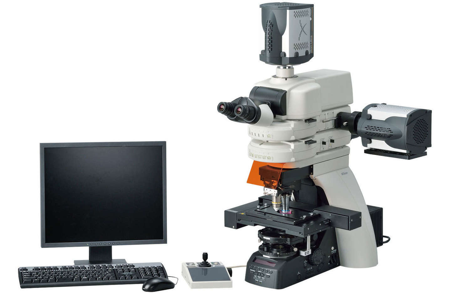
The fully motorized model Ni-E, which enables automatic observation, responds to the needs of advanced research in which control of devices such as cameras and confocal systems is linked with microscope control, and supports a wide range of cutting-edge bioscience research.
This product is available for clinical use in Europe. Please contact your local Nikon dealer for information on clinical use of this product in your country.
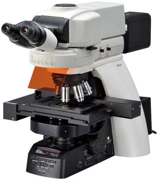
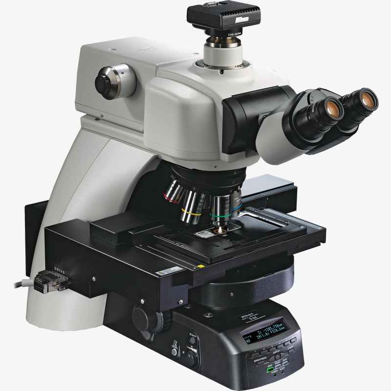
Fully motorized model equipped with motorized focus
- Motorized focus
- Broad range of motorized accessories available
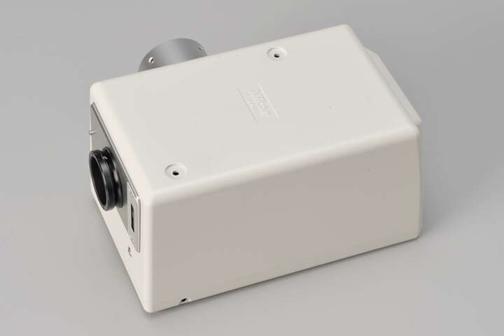
- Can be equipped with confocal systems
- Two focusing mechanism options: focusing stage and focusing nosepiece
- Ideal for advanced research
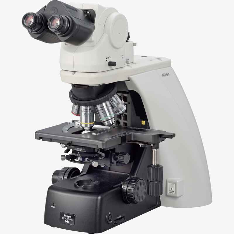
Manual model with built-in LED light source
- Manual focus
- Motorized nosepiece and motorized fluorescence cube turret available
- Built-in high color rendering LED light source
- Standard model suitable for brightfield observation of pathological specimens and digital imaging
- Supports a variety of activities, from research to routine examinations
Caractéristiques-clés
Flexible system expandability to meet diverse needs
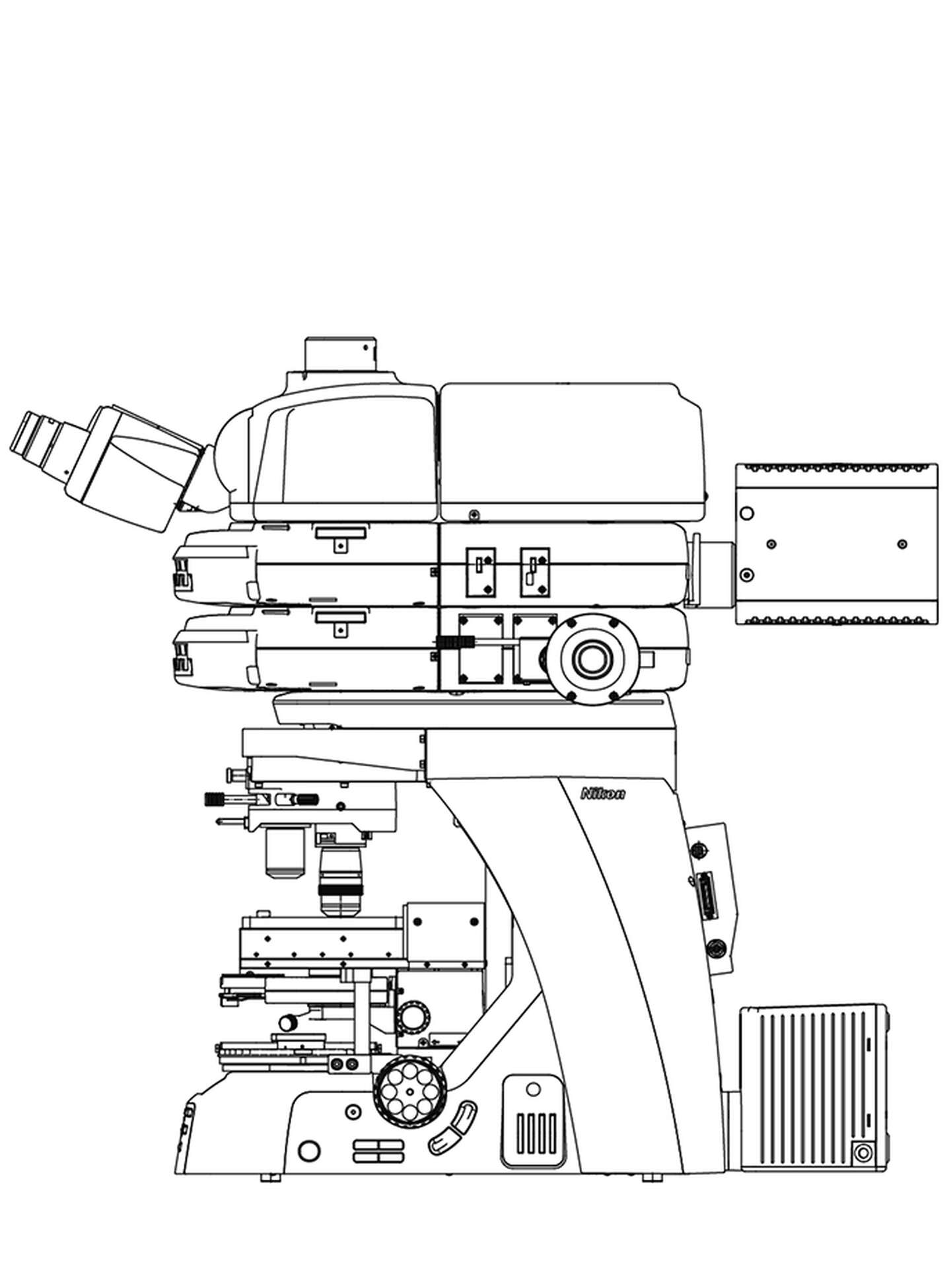
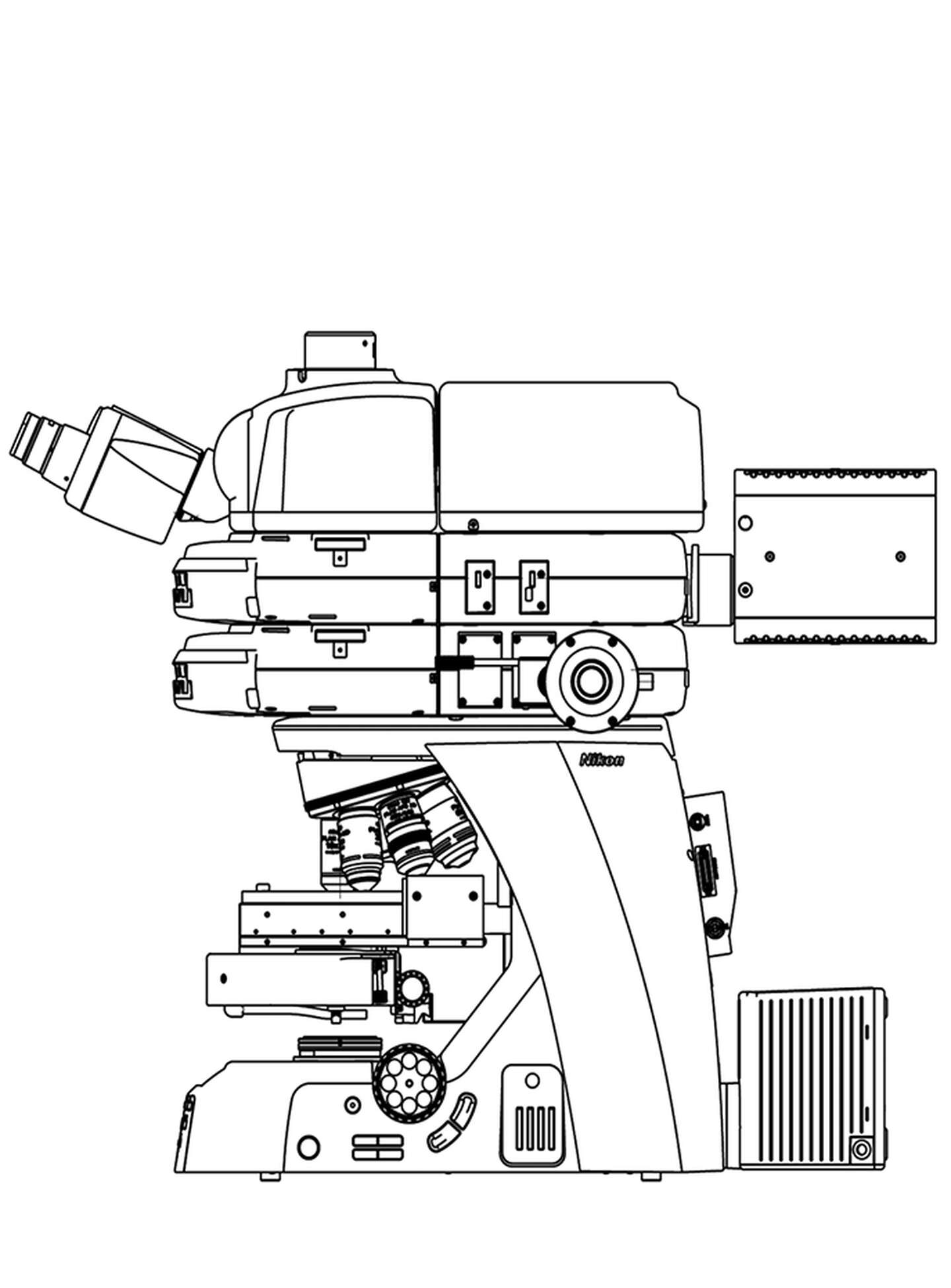
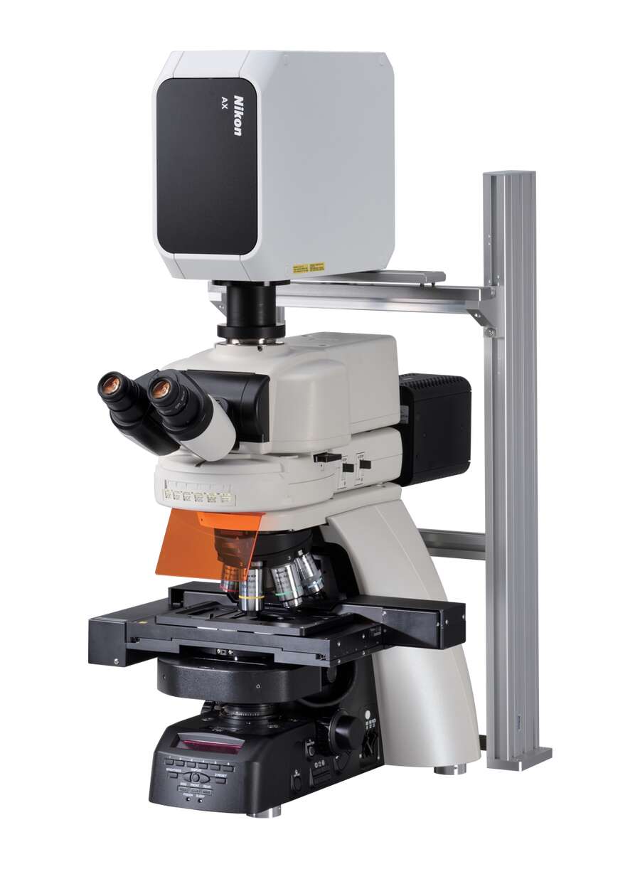
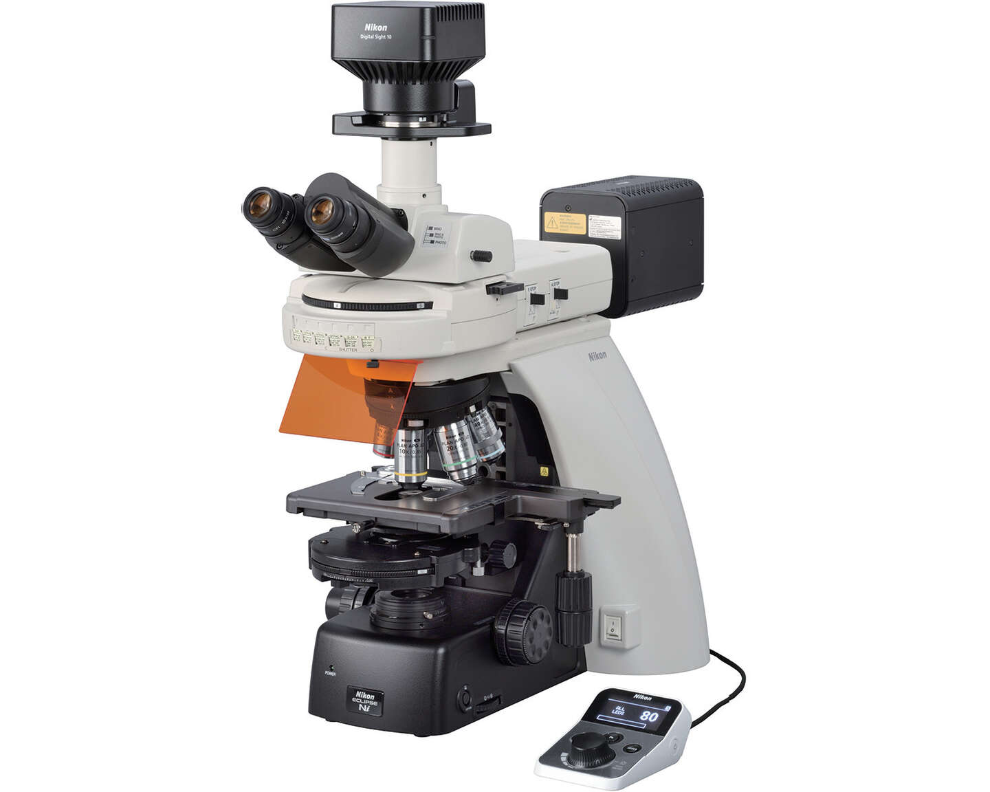
Ni-E (plate-forme de mise au point) configuré avec un illuminateur d'épifluorescence motorisé, un condensateur motorisé et un tube inclinable quadroculaire motorisé
Stratum structure capable of mounting multiple optical paths
Nikon's proprietary stratum structure enables simultaneous mounting of two optical paths on one microscope. This structure allows vertical attachment and individual control of an excitation filter cube turret and a barrier filter cube turret, enabling support for a variety of applications.
Focusing nosepiece type
Focusing stage type
Focusing mechanisms can be modified for the application
Ni-E's focusing mechanism can be selected from focusing stage type and focusing nosepiece type. The focusing nosepiece type enables a fixed-stage experimental system and meets the needs of in vivo imaging.
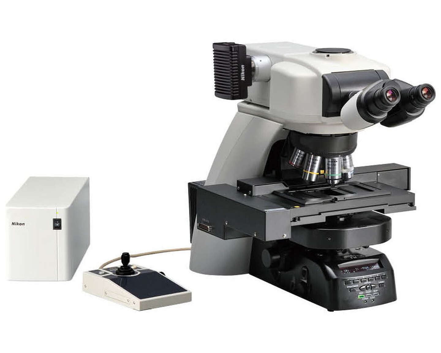
High-speed motorized accessories
The fully motorized model Ni-E can be custom-combined with a wide array of motorized accessories, including a motorized XY stage and a motorized DSC zooming port, depending on the application. The manual model Ni-L can also be equipped with motorized accessories such as the motorized nosepiece, motorized epi-fluorescence cube turret, and motorized shutter.
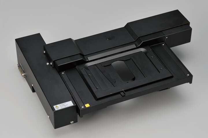
Motorized XY stage (Ni-E)
Motorized DSC zooming port (Ni-E)
Motorized epi-fluorescence cube turret (Ni-E, Ni-L)
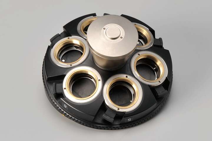
Motorized DIC sextuple nosepiece (Ni-E, Ni-L)
Supports a wide range of observation methods
Ni series accessories are segmented by function, allowing you to select required units and flexibly combine them to create lean and effective system configurations.
Confocal imaging
Configuration du type de platine de focalisation Ni-E

Configuration avec porte-objectifs de mise au point Ni-E
Combining the Ni-E’s high-precision Z-focus mechanism with Nikon’s confocal microscope system allows high-resolution, high-S/N-ratio imaging of 3D structures of organs and cells. The Ni-E has a highly stable structure suitable for mounting a confocal scanner and features a wide luminous flux that enables bright confocal imaging.
Simultaneous multichannel imaging
Ni-E (plate-forme de mise au point) configuré avec un illuminateur d'épifluorescence motorisé, un condensateur motorisé et un tube inclinable quadroculaire motorisé
The Ni’s flexible stratum structure allows the back camera port unit and the epi-fluorescence attachment to be mounted at the same time, enabling simultaneous image acquisition of two different wavelengths with each camera. This enables the capture of high-resolution images in the entire frame for each wavelength without dividing the CCD chip. The use of individual cameras for acquisition allows the user to tailor acquisition parameters for each channel independently, allowing acquisition of high-sensitivity FRET images.
* For information about compatible cameras, contact Nikon or Nikon dealers.
Imaging of fluorescent-labeled specimens
Ni-L combination
In combination with a motorized nosepiece and a motorized epi-fluorescence cube turret, each unit is automatically controlled according to the camera settings such as exposure time, camera gain, and time interval, allowing fast, efficient image acquisition.
The motorized epi-fluorescence cube turret shutter, which helps to reduce photobleaching of specimens, is easily operated with a convenient Ni-L remote control pad.
Automated imaging of pathological specimens
Ni-E combination
With the Ni-E, optimal brightness can be automatically adjusted with objective changeover, eliminating the need for manual adjustment. By controlling the optical zooming of the motorized DSC zooming port for quadrocular tube, it is possible to capture images with the desired angle of field while maintaining the image quality.
Ni-E provides fully motorized microscope control
Microscope operations related to observation can be automatically controlled as previously set. The Ni-E is a fully motorized model that is efficient for experiments requiring comprehensive control of various devices, such as photoactivation devices and confocal systems.
Automatic adjustment with objective changeover
Condenser, aperture, field diaphragm and ND filter are automatically set to optimal positions according to the objective magnification in use. In addition, XYZ stage travel distance per handle rotation and parfocal distance deviation correction are automatically adjusted. Microscope settings can also be manually adjusted.
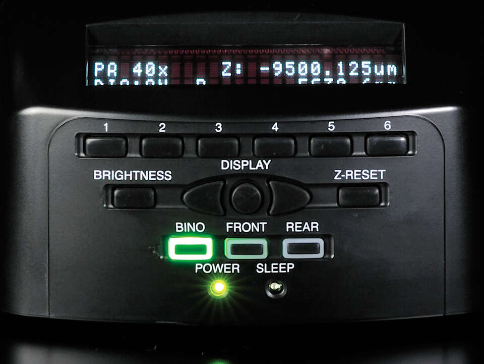
Reproducing observation conditions
Selected observation conditions can be designated to individual buttons, enabling changes to be made at the push of a button. This is particularly convenient when reproducing specific observation conditions.
High-precision motorized focusing
High-precision Z-focus provides accurate Z-position information required for use with confocal laser microscopes and supports automated Z-series acquisition. Individual coarse and fine focus knobs enhance ease of operation.
High optical performance
Nano Crystal Coat technology
Employed for the first time in microscope objectives, this anti-reflective coating consists of nanometer-size particles. It is based on semiconductor manufacturing technology and is also used for Nikon camera lenses. The coating's coarse structure and uniform arrangement of particles in a spongy construction result in extremely low reflective indices.
sans couche de nanocristal
avec couche de nanocristal


CFI Plan Apochromat Lambda D series
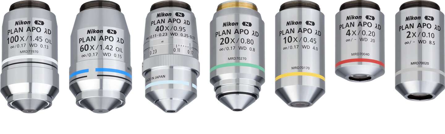
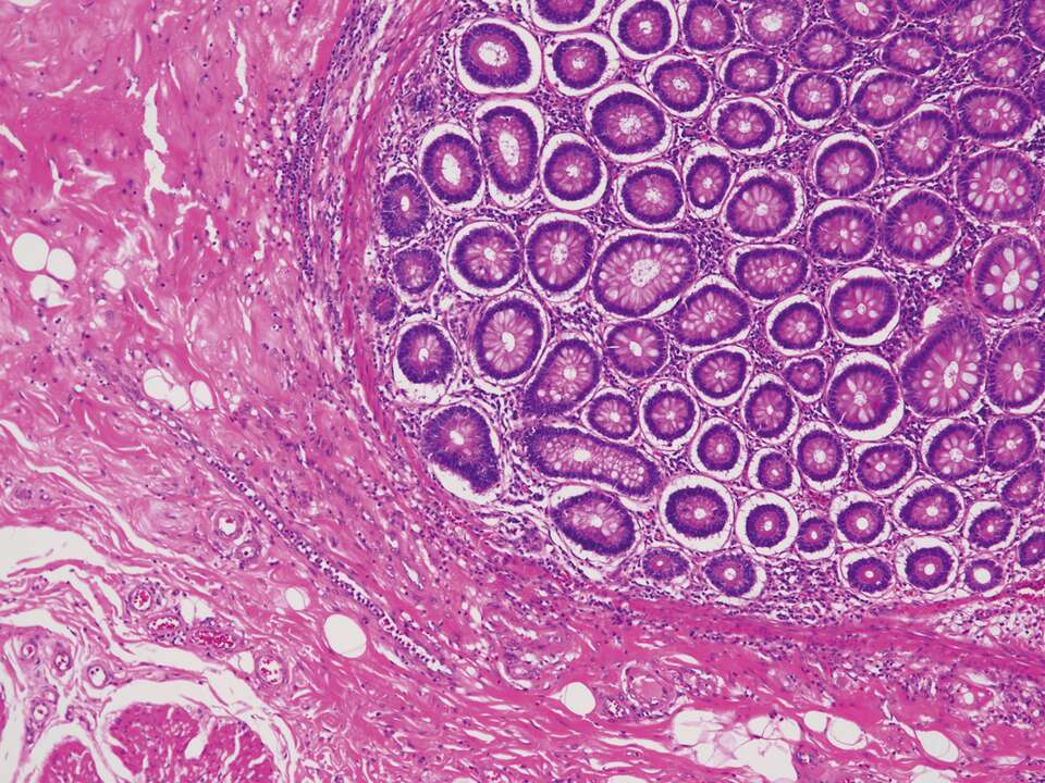
The high-refractive index glass used in the oil-immersion objectives provides uniform brightness and high image quality up to the periphery of a large 25mm diagonal field of view, enabling efficient acquisition of seamless stitched images and supports macro imaging of large samples. High transmittance and chromatic aberration correction over a wide wavelength range from 405 nm to 850 nm enable reliable quantitative data acquisition for intensity analysis of nuclear staining. These high NA lenses are ideal for brightfield and DIC observations, as well as fluorescence and confocal observations.
Water dipping objective lenses
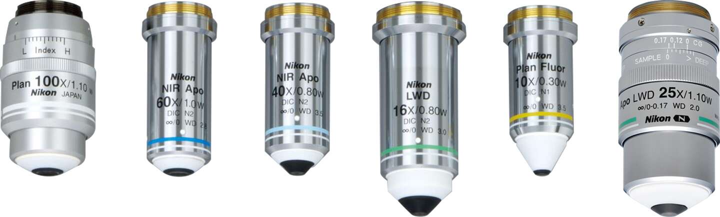
CFI Apochromat NIR 40X W/60X W objectives with long working distances and high NA provide high transmission over the near-IR wavelength range. Axial chromatic aberration is corrected up to the near-IR range, enabling high-resolution images of minute structures of thick samples during IR-DIC observations.
CFI75 Apochromat 25XC W and CFI Plan Achromat100XC W objectives featuring high NA (1.10) and long working distance (2.00 mm at 25XCW, 2.50 mm at 100XCW) are corrected for chromatic aberration in the IR range. These objectives can capture crisp images of deep regions of thick samples by adopting a mechanism to compensate for changes in spherical aberration that occur at different temperatures and observation point depths.
Uniformly bright illumination
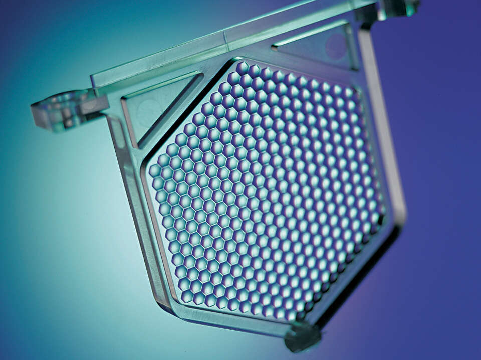
A built-in “fly-eye” lens ensures uniform and bright illumination of the field of view, from edge-to-edge, at any magnification.
Fluorescence noise elimination
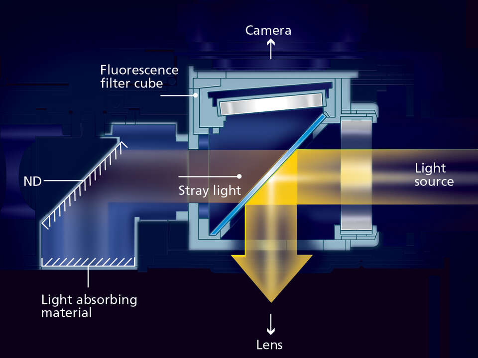
Nikon's proprietary noise terminator mechanism is employed in the epi-fluorescence cube turret and filter cubes. The S/N ratio is dramatically improved by eliminating stray light in the filter cubes, allowing images with weak fluorescent signals to be captured with high contrast and brightness.
High color rendering LED light source
The high color rendering LED light source built-into the Ni-L provides natural color reproducibility comparable to a halogen light source, as well as light uniformity, long life, and other advantages associated with LEDs, making it effective for observation of pathological specimens.
Ease of operation
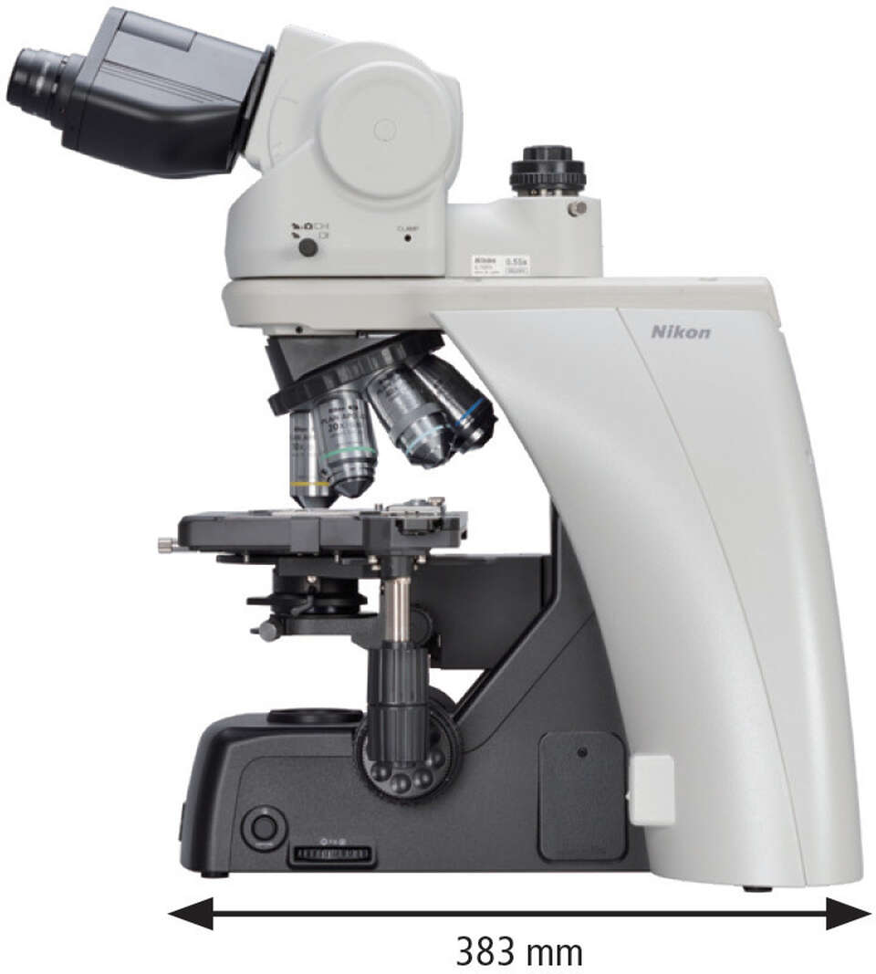
The ergonomic tube and stage handle height adjustment mechanisms allow comfortable viewing positions. The space-saving microscope base enables users to maximize their work area. A simple remote control pad for easy operation of motorized accessories is also available.
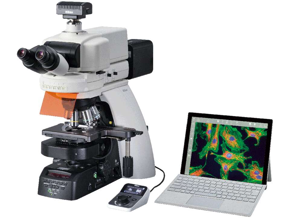
Simple digital imaging
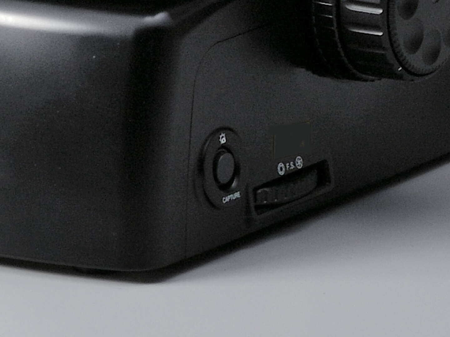
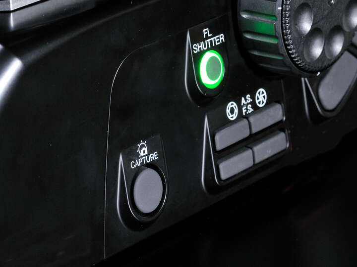
Images can be captured with Digital Sight cameras by simply pressing the image capture button located on the microscope base.
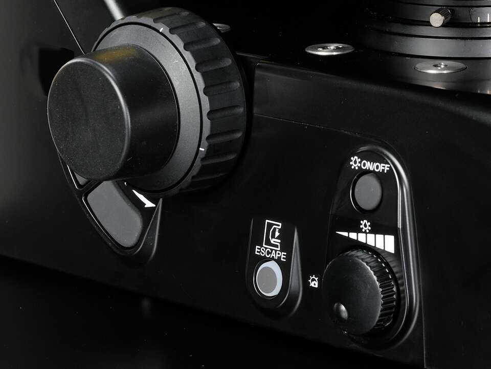
3D ergonomic design
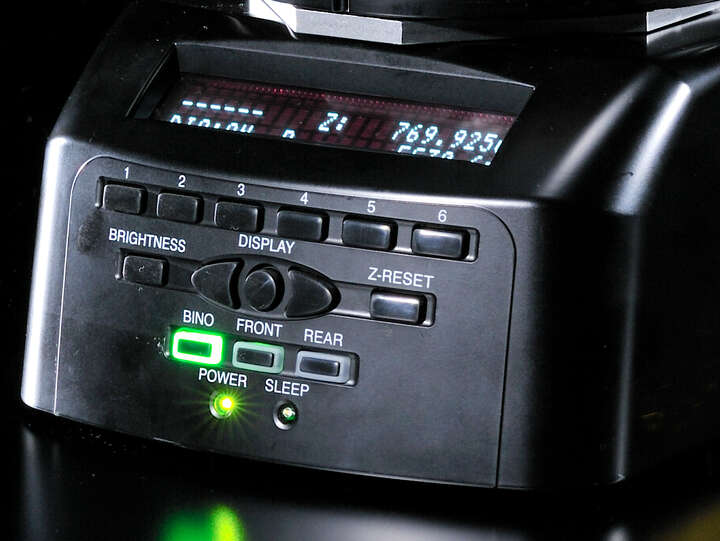
Most microscope controls can be operated with easy-to-reach buttons on the front of the Ni-E. Operation buttons on the microscope's sides are angled to allow intuitive touch-type operations during observation. Also, the microscope has a status display that can easily check the current microscope settings.
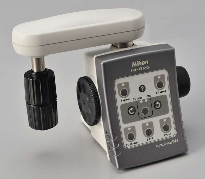
Ergo controller (Ni-E)
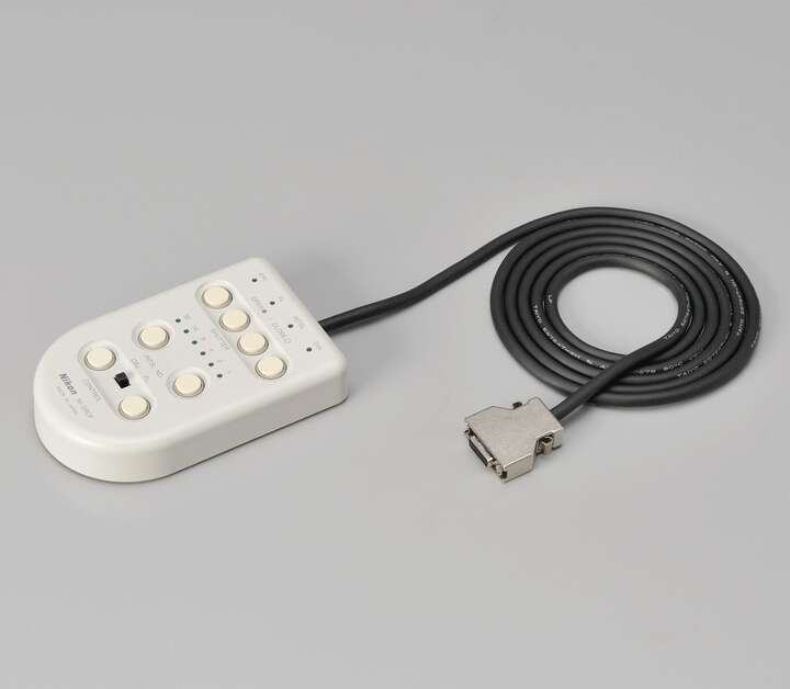
Simple remote control pad (Ni-L)
Remote controller
The simple remote control pad for Ni-L makes it easy to control the motorized accessories. An ergo-controller with similar operational feel to the actual microscope is also available for Ni-E, facilitating intuitive operation.
Compact design without a protruding lamphouse
The Ni-L has a built-in LED light source for diascopic illumination and the lamphouse does not protrude, so it is short in depth and compact, saving installation space.
Brochure de produit
Microscopes droits
- ECLIPSE Ni Series
- ECLIPSE Ci Series
- ECLIPSE FN1
- ECLIPSE Si
- ECLIPSE Ei
- ECLIPSE Ui