


Nikon Imaging Center
Northwestern University
The Nikon Imaging Center at Northwestern University Feinberg School of Medicine (NIC@NU-FSM) is an integral component of the Northwestern University Cell Imaging Facility, supported by Nikon, the Feinberg School of Medicine, Department of Cell & Molecular Biology and the Robert H. Lurie Comprehensive Cancer Center.
Established in 2008, the NIC@NU-FSM carries an important mission to:
- Augment basic research by providing access to state-of-the-art imaging equipment
- Provide training courses and organize symposia on light microscopy techniques for the benefits of the Northwestern community as well as the greater Chicago area
- Introduce cutting-edge imaging technology to Northwestern University
- Serve as a instrument evaluation and testing site for new equipment from Nikon
- Serve as a learning center for staff, researchers and students
Contact
NIC Director
email hidden; JavaScript is required
Address
Nikon Imaging CenterMorton 2-633
Feinberg School of Medicine
303 E. Chicago Avenue
Chicago, IL 60611
Website
Systems Available
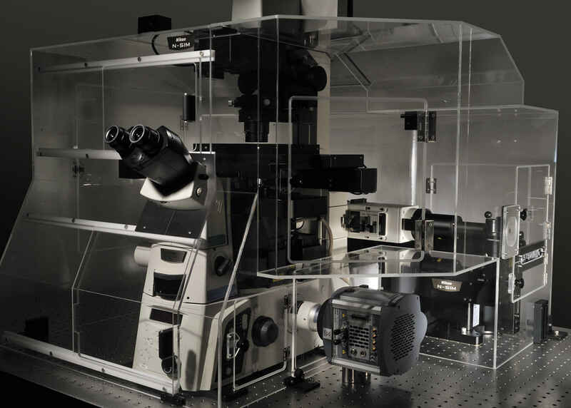
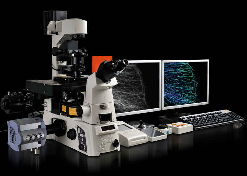
N-SIM Super-Resolution System
Featuring both Nikon's N-SIM structured illumination system and incubation, this system provides a powerful platform for live-cell super-resolution imaging.
Components
- ECLIPSE Ti-E inverted microscope with Perfect Focus System (PFS)
- N-SIM super-resolution structured illumination microscopy system
- LU5 N-SIM 4-line laser unit (405nm, 488nm, 561nm, 640nm)
- Hamamatsu ORCA-Flash4.0 V2+ sCMOS camera
- Tokai Hit stage-top incubation system
NIS-Elements software
BioStation IM-Q System
The BioStation IM-Q incorporates a microscope, an incubator and a high-sensitivity cooled CCD camera in a compact body. This all-in-one package provides a stable environment for live cells and advanced solutions for simple long-term time-lapse data acquisition.
Components
- BioStation IM-Q System
- Built-in incubation
- Phase Contrast and Fluorescence observation methods
- Built-in cooled monochrome camera
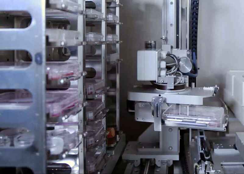
BioStation CT System
The BioStation CT combines the cell culture and imaging environments to enable accurate monitoring of individual cells and colonies while maintaining stable environmental conditions.
Components
- BioStation CT System
- Built-in incubation
- Robotic plate loader, up to 30 culture vessels
- Built-in LCD touchscreen controller

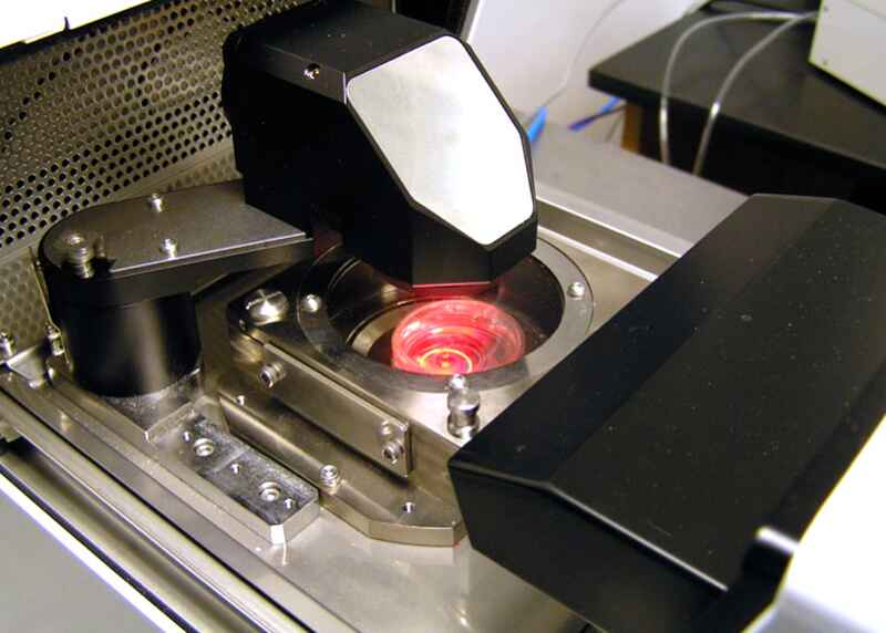
AZ-C2 Macro Confocal System
The AZ/C2 is a macro confocal microscope system which allows for the capture of confocal images allowing for better resolution of 3D specimens.
Components
- AZ100 multizoom microscope
C2 point scanning confocal system
LUN-4 4-line laser unit (408nm, 488nm, 561nm, 640nm)
- DU3 3-channel confocal detector
- Qimaging Retiga 2000 scientific CCD camera (widefield)
- Excelitas X-Cite XLED1 LED illumination system
NIS-Elements software
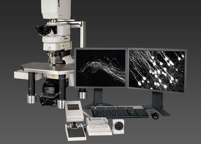
A1R MP Resonant Scanning Multiphoton System
Deep tissue imaging is made possible using this Nikon A1R MP multiphoton system configured on Ni-E upright microscope.
Components
Ni-E upright microscope
- Nikon A1R MP resonant scanning multiphoton system
- Coherent Ti:S Chameleon Vision-S laser (tunable 690-1050nm)
- Episcopic GaAsP and diascopic non-descanned detectors (NDDs)
- Tokai Hit stage-top incubation system
- Mad City Labs Nano-Z series Z-axis piezo stage
NIS-Elements software
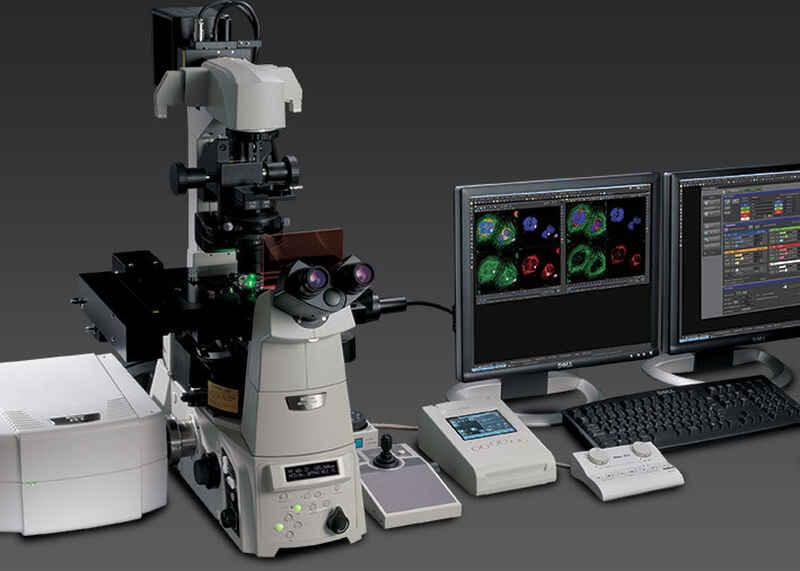
N-STORM/TIRF/CSU-X1 Super-Resolution & Spinning Disk Confocal System
This multimodal TIRF, super-resolution N-STORM, and spinning disk confocal system is adaptable to a variety of samples.
Components
- ECLIPSE Ti-E inverted microscope with Perfect Focus System (PFS)
N-STORM super-resolution single molecule localization microscopy system
Yokogawa CSU-X1 spinning disk confocal
- LUN-V 6-line laser unit (405nm, 458nm, 488nm, 515nm, 561nm, 640nm)
- Andor iXon 897 EMCCD camera
- Tokai Hit stage-top incubation system
NIS-Elements software
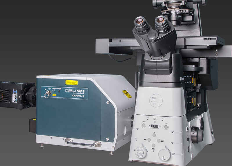
TIRF/FRAP/CSU-W1 Spinning Disk Confocal System
Combining the Nikon Ti2-E microscope with Yokogawa CSU-W1 spinning disk and emission splitting optics unlocks fast and large field of view confocal imaging.
Components
ECLIPSE Ti2-E inverted microscope with Perfect Focus System 4 (PFS4)
Yokogawa CSU-W1 spinning disk confocal
- Nikon TIRF illuminator
- Bruker Mini-Scanner galvo-based stimulation system
- LUN-V 6-line laser unit (405nm, 458nm, 488nm, 515nm, 568nm, 640nm)
- MAG Biosystems Dual-Cam two-camera emission splitter
- 2x Hamamatsu ORCA-Flash4.0 V2+ sCMOS camera (used with emission splitter)
- Photometrics Prime95B sCMOS camera
- Tokai Hit stage-top incubation system
NIS-Elements software
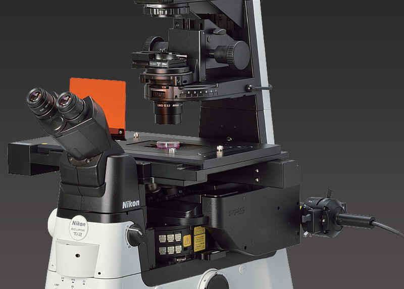
Inverted Widefield Microscope System
This fast and large field of view widefield system features a Nikon Ti2-E and Lumencor Spectra III light engine.
Components
ECLIPSE Ti2-E inverted microscope with Perfect Focus System 4 (PFS4)
Large format DS-Qi2 monochrome CMOS camera
- Lumencor SPECTRA III light engine
- Tokai Hit stage-top incubation system
NIS-Elements software
A1Rsi Resonant Scanning Confocal System
Using the Nikon A1Rsi resonant scanning confocal system with spectral detector, this system is made for linear unmixing of multiplynlabeled samples.
Components
- ECLIPSE Ti-E inverted microscope with Perfect Focus System (PFS)
- A1Rsi resonant scanning confocal system
- LU4A 6-line laser unit (405nm, 458nm, 488nm, 515nm, 561nm, 638nm)
- A1-DUS 32-channel spectral detector
- DU4G GaAsP 4-channel confocal detector
NIS-Elements software