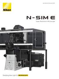
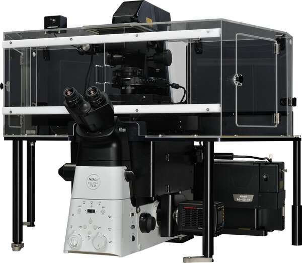
A personal super-resolution microscope that provides the same high resolution as the N-SIM S.
N-SIM E is a streamlined, affordable super-resolution system that provides double the resolution of conventional light microscopes. Combining N-SIM E and a confocal microscope allows you the flexibility to select a location in the confocal image, and easily switch to view it in super-resolution, enabling the acquisition of more detail.
Key Features
Double the resolution of conventional light microscopes
The N-SIM E utilizes Nikon's innovative approach to "structured illumination microscopy". By pairing this powerful technology with Nikon’s renowned objectives (with unparalleled NA of 1.49) the N-SIM E nearly doubles the spatial resolution of conventional light microscopes to approximately 115 nm*, and enables detailed visualization of minute intracellular structures and their interactions.
* This value is measured FWHM of 100 nm beads exited with 488 nm laser in 3D-SIM mode.
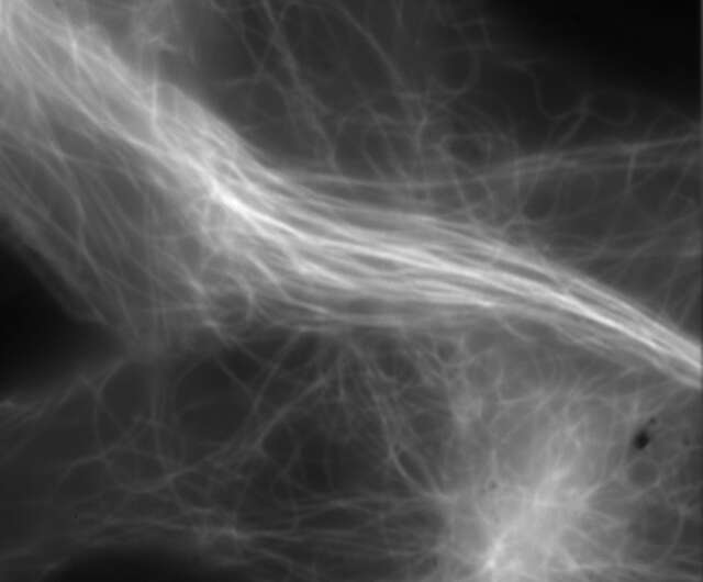
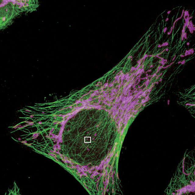
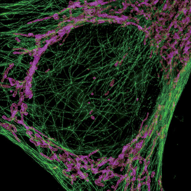
Super-resolution image (3D-SIM)
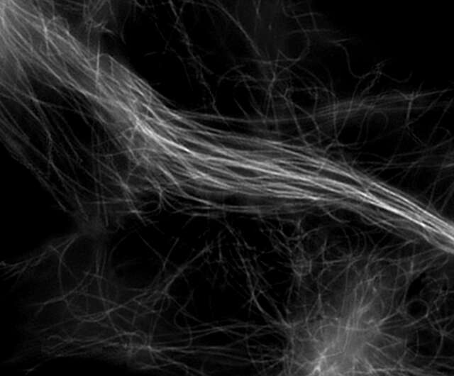
Conventional widefield image
Microtubules in B16 melanoma cell labeled with YFP
Objective: CFI Apochromat TIRF 100XC Oil (NA 1.49)
Image capturing speed: approximately 1.8 sec/frame (movie)
Reconstruction method: Slice
Photographed with the cooperation of: Dr. Yasushi Okada, Laboratory for Cell Polarity Regulation, Quantitative Biology Center, RIKEN
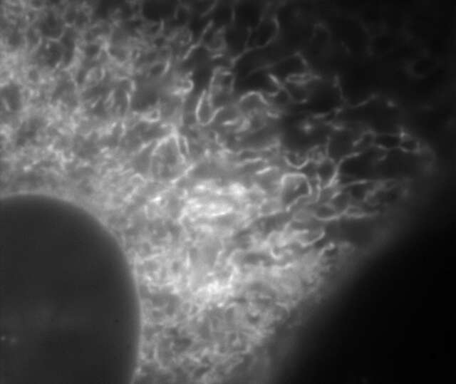
Super-resolution image (3D-SIM)
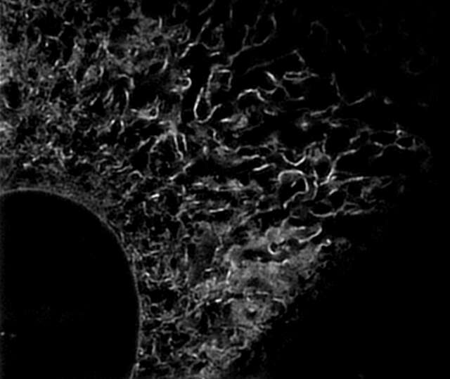
Conventional widefield image
Endoplasmic reticulum (ER) in living HeLa cell labeled with GFP
Objective: CFI Apochromat TIRF 100XC Oil (NA 1.49)
Image capturing speed: approximately 1.5 sec/frame (movie)
Reconstruction method: Slice
Photographed with the cooperation of: Dr. Ikuo Wada, Institute of Biomedical Sciences, Fukushima Medical University School of Medicine
Fast 1 sec/frame temporal resolution for super resolution imaging
N-SIM E provides fast imaging performance for Structured Illumination techniques, with a time resolution of approximately 1 sec/frame, effective for live-cell imaging.
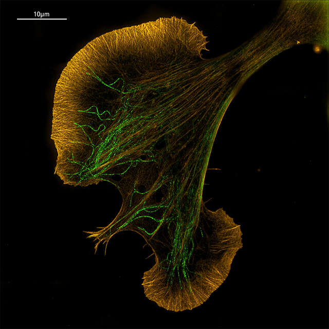
Acquire larger fields of view
N-SIM E can acquire super-resolution images with a large field of view of 66 µm square. This larger imaging area enables very high throughput for applications/samples that benefit from larger fields of view, such as neurons, reducing the amount of time and effort required to obtain data.
Reconstructed image size: 1024 x 1024 pixels (33 μm x 33 μm with a 100X objective)
Reconstructed image size: 2048 x 2048 pixels (66 μm x 66 μm with a 100X objective)
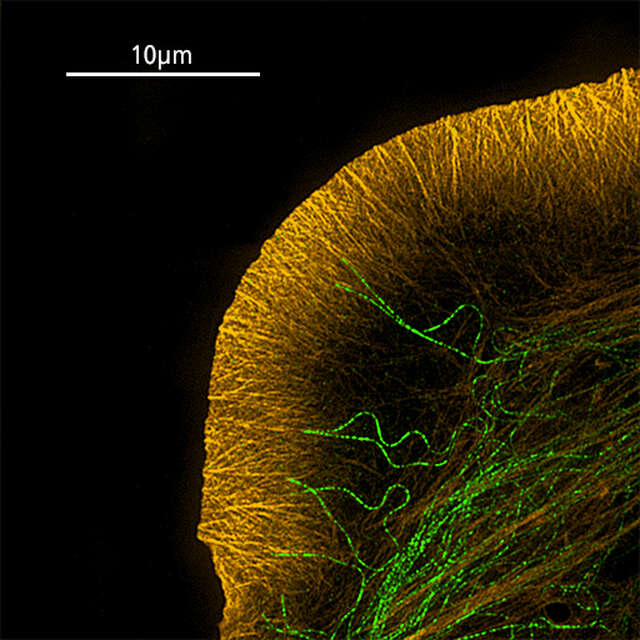
Growth cone of NG108 cell labelled with TRITC-phalloidin (F-actin, orange) and Alexa Fluor® 488 (microtubules, green)
Sample courtesy of: Dr. Shizuha Ishiyama and Dr. Kaoru Katoh, The National Institute of Advanced Industrial Science and Technology (AIST)
Axial super-resolution with 3D-SIM mode
The 3D-SIM mode generates structured illumination patterns in three dimensions to deliver a two-fold improvement in lateral and axial resolutions. Two reconstruction methods (“slice” and “stack”) are available to optimize results according to application requirements (e.g. sample thickness, speed, etc.). Slice reconstruction is suitable for imaging living cells at specific depths, as it allows axial super-resolution imaging with optical sectioning at 300 nm resolution. Optional stack reconstruction, based on Gustafsson’s theory, is suitable for acquisition of volume data as it can image thicker specimens with higher contrast than slice reconstruction.
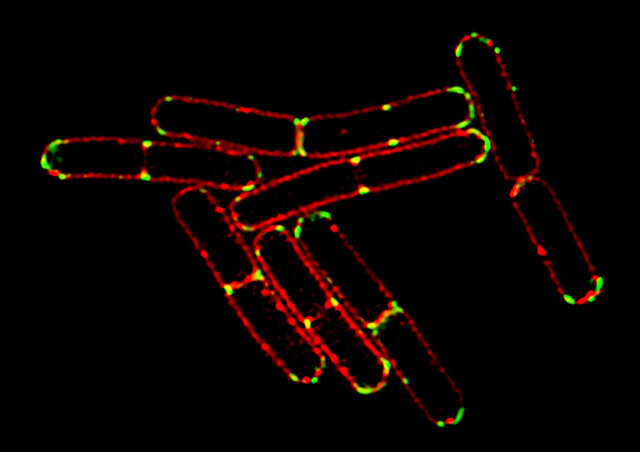
3D-SIM image
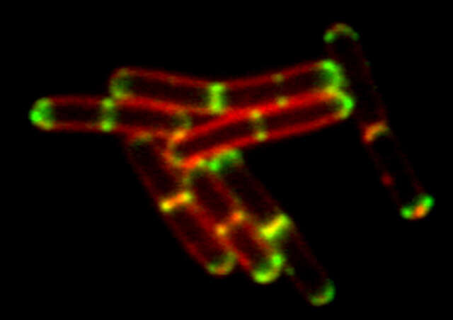
Conventional widefield image
Bacillus subtilis bacterium stained with membrane dye Nile Red (red), and expressing the cell division protein DivIVA fused to GFP (green).
The super-resolution microscope allows for accurate localization of the protein during division.
Reconstruction method: Slice
Photos courtesy of: Drs. Henrik Strahl and Leendert Hamoen, Centre for Bacterial Cell Biology, Newcastle University
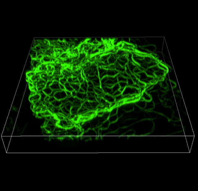
3D-SIM (Volume view)
Width: 26.16 μm, Height: 27.11 μm, Depth: 3.36 μm
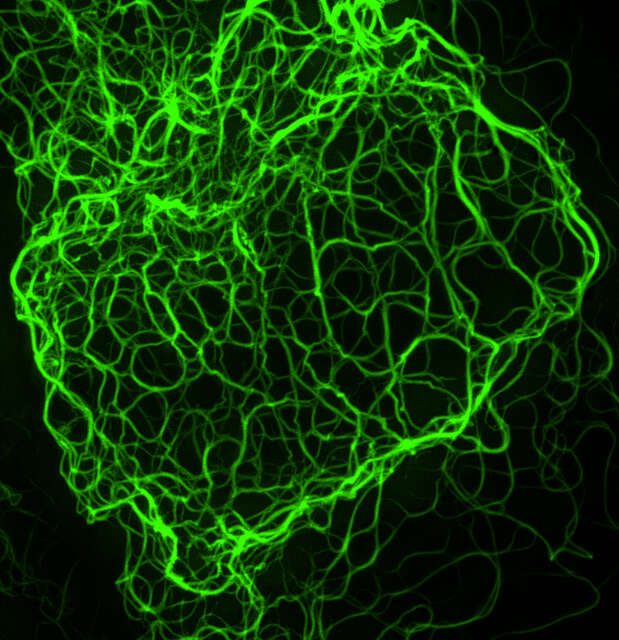
3D-SIM (Maximum projection)
Mouse keratinocyte indirectly immunolabeled for keratin intermediate filaments and visualized with Alexa Fluor® 488 conjugated secondary antibodies.
Reconstruction method: Stack
Photo courtesy of: Dr. Reinhard Windoffer, RWTH Aachen University
Seamless switching between imaging modalities for multi-scale experiments
The N-SIM E can be combined with a confocal microscope such as the AX/AX R. A desired location in a sample can be specified in a low-magnification/large FOV confocal image and acquired in super-resolution by simply switching the imaging method. Combining a confocal microscope with a super-resolution system can provide a method for gaining larger contextual views of super-resolution information.
Select the location to acquire a SIM image in a confocal image
Acquire the SIM image of the selected location
3-color multi-laser super-resolution capability
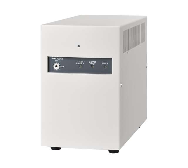
The compact LU-N3-SIM laser unit dedicated for N-SIM E is installed with the three most commonly used wavelength lasers (488/561/640), enabling super-high resolution imaging in multiple colors. It enables the study of dynamic interactions of multiple proteins of interest at the molecular level.
Objectives for super-resolution microscopes
Silicone immersion objectives
Silicone immersion objectives use high viscosity silicone oil with a refractive index close to that of live cells as an immersion liquid. Because of this improved refractive index compatibility, these objectives can provide improved photon collection capability and resolution when performing super-resolution imaging deeper into the specimen. They exhibit superior chromatic aberration correction and high transmittance over a broad range of wavelengths.
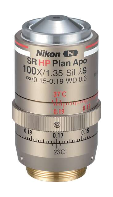
CFI SR HP Plan Apochromat Lambda S 100XC Sil
Mouse brain section labeled with tdTomato expressing neurons
Immersion objectives
The system can be configured with either a 100X oil immersion type, which is suitable for the imaging of fixed samples, or a 60X water immersion type, which is optimal for time-lapse live-cell imaging. The SR objectives are aligned and inspected using wavefront aberration measurement technologies to ensure the lowest possible asymmetric aberration and the superb optical performance required for super-resolution imaging.
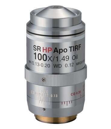
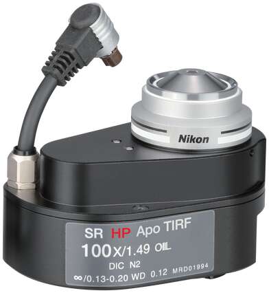
CFI SR HP Apochromat TIRF 100XC Oil
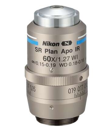
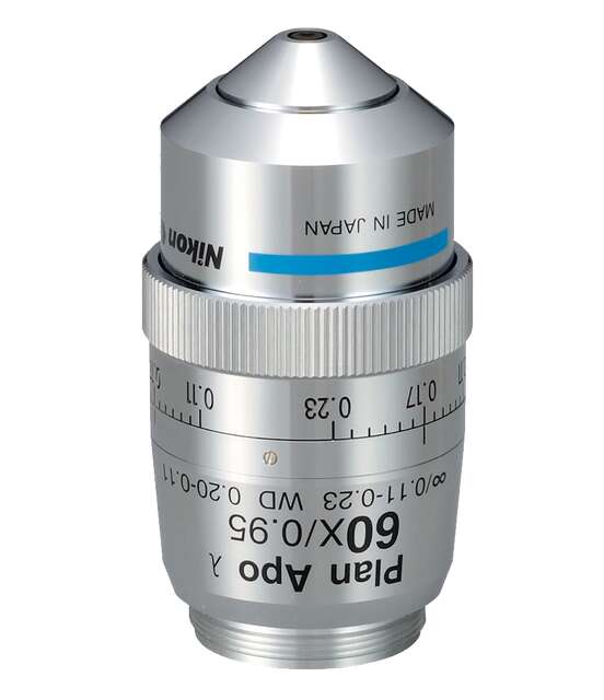
CFI SR Plan Apochromat IR 60XC WI
CFI SR HP Apochromat TIRF 100XAC Oil
Dry objective
The N-SIM E is compatible with dry objectives, making both super-resolution imaging and confocal imaging available without switching lenses. Low-magnification, wide field-of-view dry lenses enable high resolution observation even at the periphery of sample tissues.
* Dry objectives support slice reconstruction

Product Brochure