Application Notes
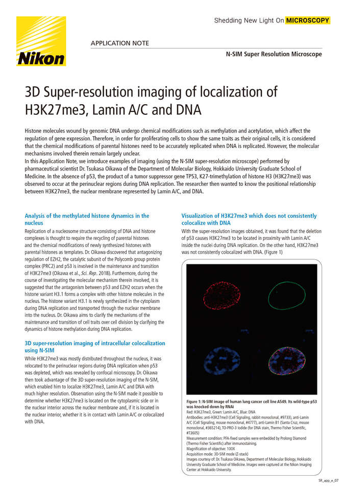
3D Super-resolution imaging of localization of H3K27me3, Lamin AC and DNA
October 2020
In this Application Note, we introduce examples of imaging (using the N-SIM super-resolution microscope) performed by pharmaceutical scientist Dr. Tsukasa Oikawa of the Department of Molecular Biology, Hokkaido University Graduate School of Medicine. In the absence of p53, the product of a tumor suppressor gene TP53, K27-trimethylation of histone H3 (H3K27me3) was observed to occur at the perinuclear regions during DNA replication. The researcher then wanted to know the positional relationship between H3K27me3, the nuclear membrane represented by Lamin A/C, and DNA.
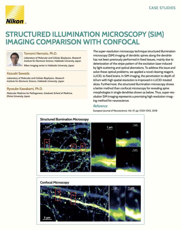
Structured Illumination Microscopy (SIM) Imaging Comparison with Confocal
September 2018
The super-resolution microscopy technique structured illumination microscopy (SIM) imaging of dendritic spines along the dendrite has not been previously performed in fixed tissues, mainly due to deterioration of the stripe pattern of the excitation laser induced by light scattering and optical aberrations.
N-SIM for Quantitative Ultra-Structural Analyses of the Nuclear Lamina
October 2016
Super-resolution Structured Illumination Microscopy (SIM), available from Nikon via the N-SIM S and N-SIM E systems, allows for the observation of details inaccessible to traditional microscopes, such as confocal and widefield. In this application note we see how the N-SIM system enables quantitative multi-color evaluation of the distribution of different nuclear lamin proteins and the structures they form.
Product Brochure