


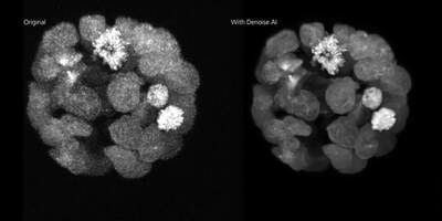
Image courtesy of Laurence Pelletier Lab, LTRI
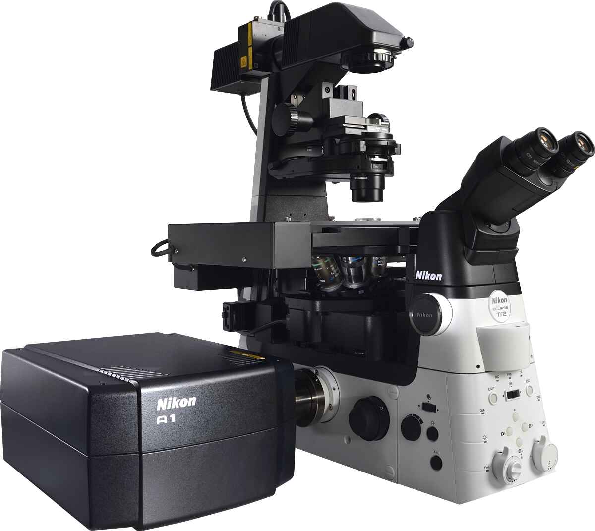
A1 HD25 / A1R HD25
Confocal Microscope System
Discontinued Replaced by AX / AX R with NSPARC
A confocal microscope that captures images with a 25 mm field of view, nearly twice the area of conventional point scanners.
Capturing images of large samples such as tissues, organs and live model organisms requires both extending the detectable area of cellular responses and increasing image capture speed. The A1 HD25/A1R HD25 confocal microscope has the largest field of view (25 mm) in its field, enabling users to expand the limits of scientific research.

Recommended Model:
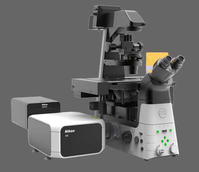
AX / AX R with NSPARC
Nikon’s latest confocal microscope, leveraging deep learning-based AI tools to simplify acquisition and improve image quality. Fast scanning combined with a large 25 mm field of view enables high throughput imaging with unmatched edge-to-edge image quality. Super-resolution provided by the NSPARC detector.