Focus sulla virologia
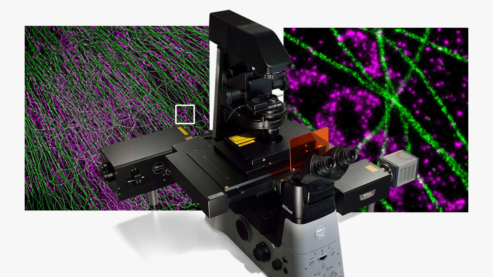
Le dimensioni estremamente piccole dei virus (5-300 nm) rendono difficile lo studio della la loro struttura e funzione. Negli ultimi secoli sono stati scoperti molti virus e sono stati persino sviluppati molti vaccini contro di loro, ma è stata l'invenzione del microscopio elettronico nel 1931 a illuminare per la prima volta la loro struttura complessa. Da quel momento, le tecniche di microscopia ottica come l'acquisizione di immagini a fluorescenza sono arrivate a svolgere un ruolo importante nella ricerca sui virus, consentendo di studiarne l'attività nei sistemi viventi. Nikon è un produttore leader di sistemi avanzati di microscopia ottica per l'acquisizione di immagini dell'attività dei virus in tempo reale ad alta risoluzione e alto rendimento. Data la crescente importanza di analisi rapide e affidabili dell'attività dei virus, Nikon è più impegnata che mai nello sviluppo e nel supporto di sistemi avanzati per questo tipo di lavoro.
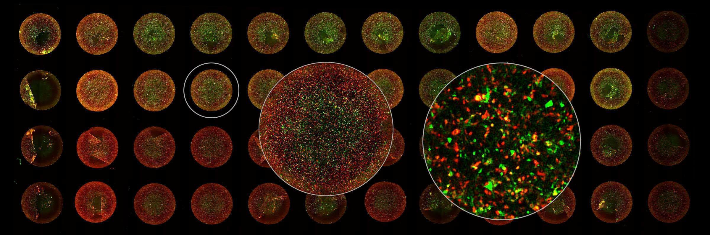
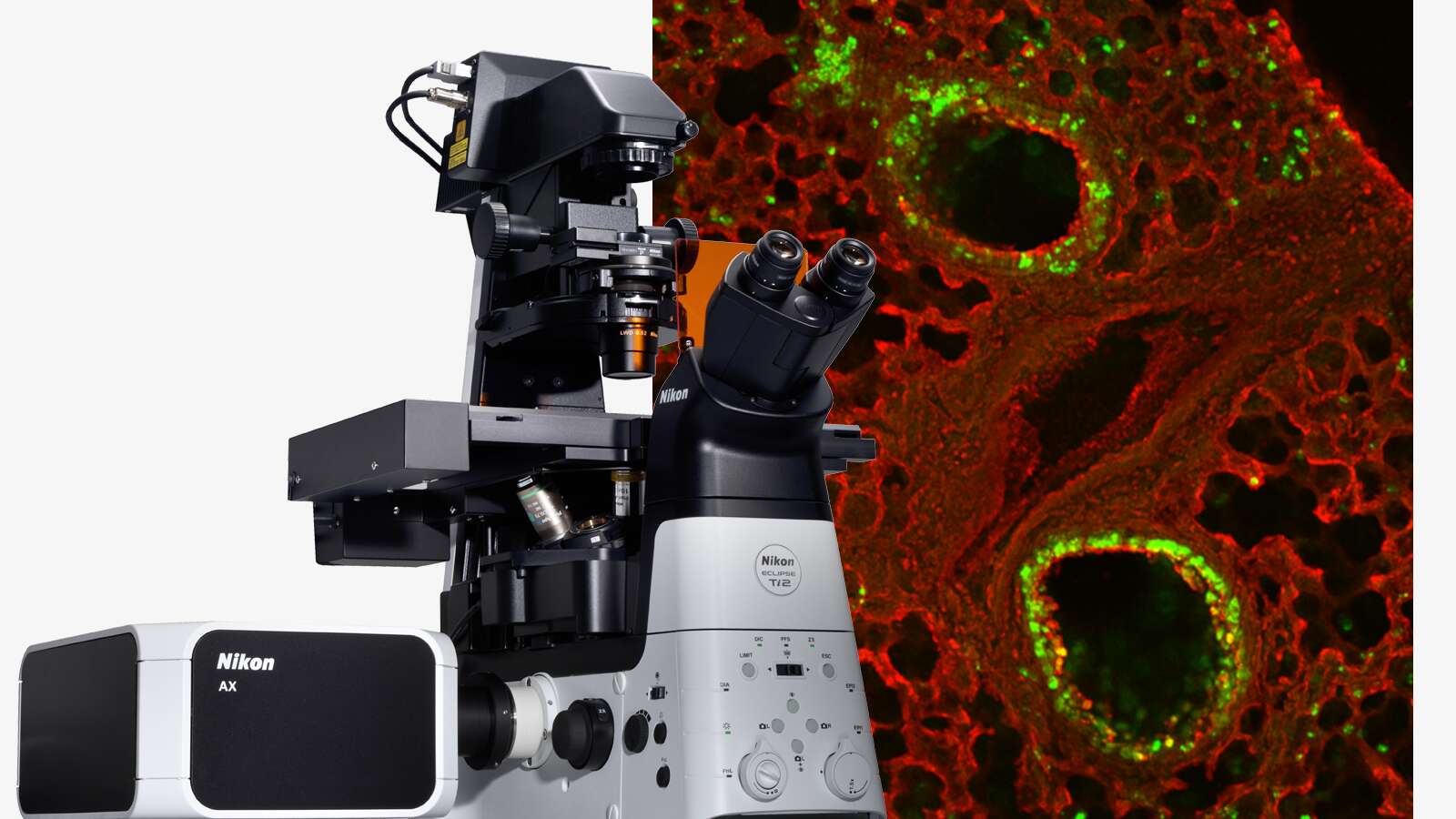
Image courtesy of Rudolph Reimer, Heinrich Pette Institute, Leibniz Institute for Experimental Virology